Introduction to Neuroanatomy - Part 3 (Brain Structures - Telencephalon and Diencephalon)
Brain Structures and their Functions
To learn more about neuroanatomy, please check out: "Neuroanatomy Text and Atlas," by John H. Martin.
In the last post, we ended explaining the development of the spinal cord. We will continue this discussion by exploring the structures of the brain from a bottom-up perspective and describe their functions.
Cranial and Non-cranial Nerves
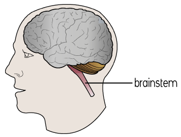
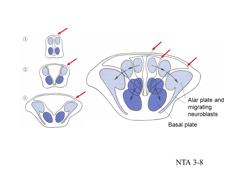 If you read the last post (Here is the link: http://hbookreviews.blogspot.com/), you should remember that the spinal cord was divided by two plates: basal and alar. When we move up from the spinal cord into the brain we would observe a collection of structures known as the brainstem. In here, the plates become nerves. The alar plate becomes the cranial nerve sensory nuclei and the basal becomes the cranial nerve motor nuclei. Remember that a nerve is a bundle of axons in the central nervous system (CNS) and that nuclei are a collection of cell bodies that are also located in the CNS. Thus, the phrase "cranial nerve motor nuclei" tells us what the body part is (nerve=bundle of axons (1)), where it is (the nuclei is located inside the CNS, which is either the brain or the spinal cord (2)), and what it does (motor refers to sending information across the spinal cord that will eventually create movement. As you can see from the picture to the right, the dorsal or alar plates moved laterally in the brainstem.
 The brain stem is composed of three structures. They are the medulla, the pons, and the midbrain (3). Before we go into their functions, I want to keep talking about nerves. Each of these structures has non-cranial nerve nuclei. The inferior olivary nuclei are located in the medulla and they are responsible for hearing functions. The pontine nucleus is located in the pons and it is responsible for skilled movement control. The red nucleus and the substantia nigra are located in the midbrain. They are responsible for descending motor and ascending motor information, respectively. We will cover more of the brain stem later.
 There is a total of twelve cranial nerves. Next to them I'll write their functions. They are:
- Olfactory - This nerve helps with the sense smell (6).
- Optic - This nerve helps with vision by transmitting information from the retina to the brain (5).
- Oculomotor - This nerve helps with eye movement and accommodation, as well as pupil constriction (4).
- Trochlear - This nerve helps the eye move up and down (7).
- Trigeminal - This nerve sends somatosensory (this means sensory information from the skin) information from the face and movement of the jaw (8).
- Abducens - This nerve helps with side to side movement of the eye (9).
- Facial - This nerve helps send somatosensory information from the ear and sensory information from the tongue. As well as, movements of the face (facial expressions) (10).
- Vestibulocochlear - This is a sensory nerve that deals with audition and balance (11).
- Glossopharyngeal - This nerve sends somatosensory information from the tongue and the pharynx. In addition, it sends sensory (taste) information from the posterior one-third of the tongue (12).
- Vagus - This nerve has some motor and sensory functions that range from swallowing and gland control to taste and involuntary muscle movement in the viscera (13).
- Accessory - This nerve helps with the movement of the head (14).
- Hypoglossal - This nerve helps with the movement of the tongue (15).
Prosencephalon

Now, that we covered the twelve cranial nerves, we are going to explore the structures of the brain using an embryological perspective. Do you remember the three main divisions? (It was covered in the second post) They were the prosencephalon, mesencephalon, and rhombencephalon. The former was two subdivision, they are the telencephalon and the diencephalon. The first one can be divided into three smaller parts: the cerebral cortex, the basal ganglia, and the limbic system, which is made up of the amygdala and the hippocampus. I know.. I know... It may seem as if it is too much information to process at once, but we will break it down apart in order to make it easier to understand. Think of the telencephalon as the superior part of the brain, as the cerebral hemispheres. The picture to the right also includes the corpus callosum in the telencephalon. This is the structure that connects both hemispheres, it helps by letting them communicate with each other (It is Latin for callous body).

To understand the function of the cerebral cortex, we have to explore first the lobes of the brain. As you can see there are four major lobes, they are the frontal, parietal, temporal, and occipital lobes. The first lobe is in charge of a variety of functions (16). The prefrontal cortex is located in the most rostral part of the frontal lobe. It is in charge of executive function, which is a set of cognitive skills that include planning, inhibition, and logical thinking. The frontal lobe also handles movement. There is a fissure that separates the frontal and parietal lobe is called the central fissure. The gyrus before the central fissure is called the precentral gyrus and it is where the majority of motor tasks are processed. Before the precentral gyrus, there is a structure known as Broca's area. This is the location where speech is produced. The parietal lobe, which is located after the central sulcus or fissure, handles somatosensory information (16). The postcentral gyrus is where the primary somatosensory cortex is located. This means that it handles sensory info from the skin such as pain, pressure, and temperature. The occipital lobe is where the primary visual cortex is located. Primary auditory cortex is located in the temporal lobe. In this lobe, there is a structure known as Wernicke's area. This is the opposite of Broca's area because it handles the understanding of speech. It is important to note that once sensory information arrives at its primary location in the cortex, it then goes to its respective association cortex where it undergoes further processing (17). In addition, the cortex has two divisions. They are neocortex and allocortex. The former is the most recent cortex in terms of evolution and it helps with higher cognitive functions (18). The latter is the older cortex and deals with more primitive functions. It has two subdivisions: archicortex and paleocortex. Both of them develop "in association with the olfactory system" and it doesn't have a layered structure (19).
 That was the first part of the telencephalon, the second structure would be the basal ganglia. Remember that ganglia are collections of cell bodies outside of the central nervous system (CNS), however, the basal ganglia is an exception because it is located in CNS. It is one of the structures that deal with movement. The structure that deals with memory looks like a seahorse that is why its name is hippocampus (latin for seahorse (20)). Close to the hippocampus is the amygdala (amygdala is latin for almond, it was given this name because of its shape), which is the structure that is responsible for emotions (21). In summary, the telencephalon is made up of the cerebral cortex, the basal ganglia and the limbic system, which includes the hippocampus and amygdala
 The second subdivision of the prosencephalon is the diencephalon. This subdivision is made up of several structures. One of them is the thalamus, which is a structure made up of nuclei that are responsible for sending sensory information to its respective cortex (22). Another important structure in the diencephalon would be the hypothalamus. This is responsible for the four F's: fighting, fleeing, feeding, and fornicating. As you can see this part of the brain is very primitive from an evolutionary perspective because of it functions. The epithalamus is also located in the diencephalon and it is made up of the habenula and the pineal gland (23). The former is involved in motivation and reward (24) and the latter is not where the mind and body interact like Rene Descartes used to believe, but rather it is responsible for segregating melatonin and it regulates the circadian rhythm (25).
C-Shaped Structures
During development, the telencephalon undergoes an incredible expansion in terms of neurogenesis. On average, there are about 250, 000 neurons being developed per minute (22). As a result of this, structures start to develop a c-shape. One of the structures that has this shape is the cortex, which includes the four lobes that were covered before. Other structures include the ventricles, which are cavities that contain cerebrospinal fluid (22), the corpus callosum, which connects the two hemispheres, and the caudate nucleus.
References
1. http://www.dictionary.com/browse/nerve.
2. http://www.med.umich.edu/lrc/coursepages/m1/anatomy2010/html/modules/NS_overview_module/NS_Overview_06.html
3. https://www.britannica.com/science/brainstem
4. http://medical-dictionary.thefreedictionary.com/oculomotor+nerve
5. https://www.sciencedaily.com/terms/optic_nerve.htm
6. http://medical-dictionary.thefreedictionary.com/olfactory+nerve
7. http://www.medicinenet.com/script/main/art.asp?articlekey=7603
8. http://www.ncbi.nlm.nih.gov/books/NBK384/
9. http://www.meddean.luc.edu/lumen/meded/grossanatomy/h_n/cn/cn1/cn6.htm
10. http://www.meddean.luc.edu/lumen/meded/grossanatomy/h_n/cn/cn1/cn7.htm
11. http://www.meddean.luc.edu/lumen/meded/grossanatomy/h_n/cn/cn1/cn8.htm
12. http://www.meddean.luc.edu/lumen/meded/grossanatomy/h_n/cn/cn1/cn9.htm
13. http://emedicine.medscape.com/article/1875813-overview.
14. http://www.ncbi.nlm.nih.gov/books/NBK387/
15. http://www.ncbi.nlm.nih.gov/books/NBK388/
16. https://www.dartmouth.edu/~rswenson/NeuroSci/chapter_11.html
17. http://www.ncbi.nlm.nih.gov/books/NBK11109/
18. http://www.dictionary.com/browse/neocortex
19. http://medical-dictionary.thefreedictionary.com/archicortex
20. https://www.britannica.com/science/hippocampus
21. http://www.dictionary.com/browse/amygdala.
22. "Neuroanatomy text and atlas," by John H. Martin.
23. http://medical-dictionary.thefreedictionary.com/epithalamus
24. https://www.hindawi.com/journals/aneu/2014/862048/
25. http://www.ncbi.nlm.nih.gov/pubmed/15589268


Your blog is big and there is a lot of good information! Thanks
ReplyDeletehttps://blog.mindvalley.com/diencephalon/
You're welcome! We are happy you have found it helpful!
Delete