Introduction to Neuroanatomy - Part 2 (The Brain and the Spinal Cord)
The Brain and the Spinal Cord
To learn more about neuroanatomy, please check out: "Neuroanatomy Text and Atlas," by John H. Martin.
Glial Cells
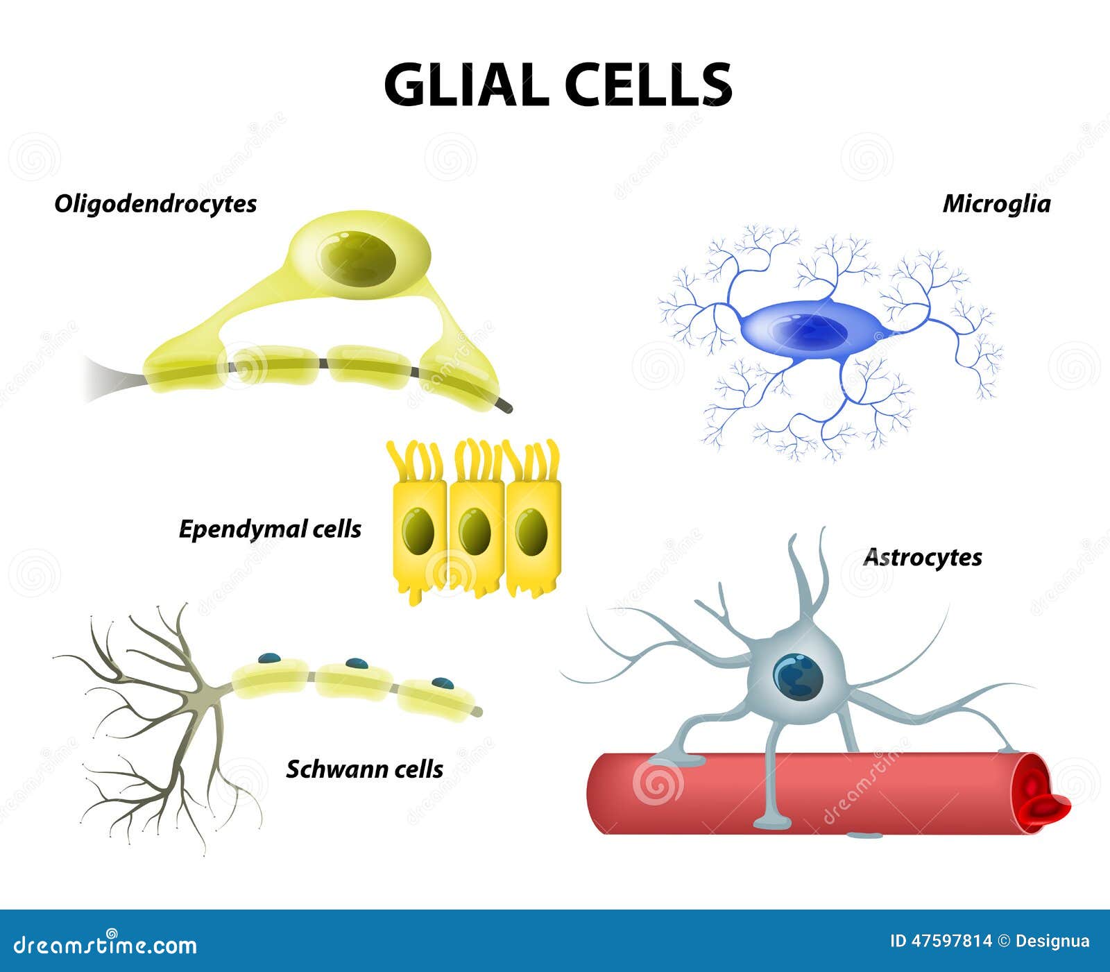 In the last cover, we covered primarily neurons, but the glial cells are also very important in the nervous system. Something important to note is that the same parts in the central nervous system (CNS) and the peripheral nervous system (PNS) have different names. We will see an example of this with a specialized type of glia cell. They outnumber neurons, however, the exact number is still being debated. One textbook affirms that the ratio is ten to one(1), while another one asserts that it is three to one (2). Whether is one or the other, the fact that there is more glial cells remains true. This might suggest that their function is vital for the nervous system. There are two types of glial cells: Microglia and macroglia. The former acts as immune cells because if there is an infection or damage detected in the central nervous system, this subdivision is in charge of phagocytizing (this means destroying a cell (3), remember that phago- means consuming (4), and cyte- means cell(5)). The latter is a group of cells that provide support or aid neurons with nutrition (1). For example, neurons have something known as the myelin sheath. This helps action potentials reach the end of the neuron in a faster manner and there are some long neurons that without the myelin sheath the action potential would never reach the terminal buttons. The cells that provide the myelin sheath to neurons are macroglia. Remember how we mentioned that the same parts have different names depending whether they are in the central nervous system (CNS) or the peripheral (PNS) one? Well, the cells that myelinate axons in the PNS are called Schwann cells and in the CNS they are called oligodendrocytes. Both of these cells
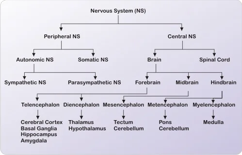
The Development of the Central Nervous System
If you saw carefully the graph above, you may have noticed that there are certain subdivisions in the brain: the telencephalon, etc. These will be covered in detail in this section, where we will cover the development of the CNS.

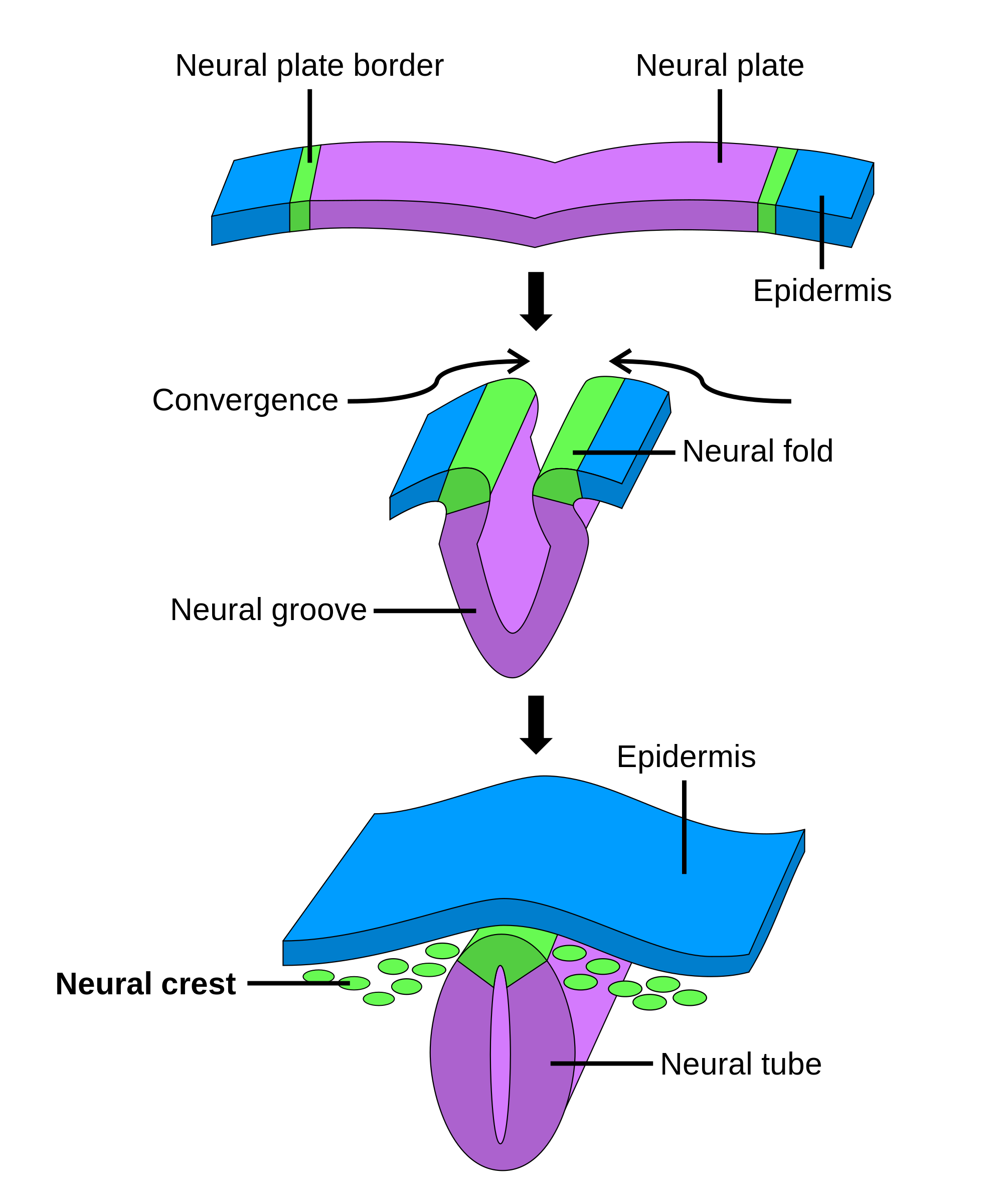 The embryo is composed of three layers: the endoderm, the mesoderm, and the ectoderm. They are the inner, middle, and external layers, respectively, in the embryo. The neural plate is a part of the ectoderm that later develops into the neural tube and the neural crest (13). The cells from the latter move to eventually create different types of cells such as neurons and glial; it is important to note that wherever they go will affect the type of cell they will become (14). The neural tube will develop into the brain and the spinal cord (15). The neural plate folds itself in order to create the neural tube. There are several points of view in order to study the brain in terms of orientation. The first one is rostral, which refers to a frontal view, caudate, which refers to a rear perspective, dorsal, which refers to looking at things from above, and ventral, which refers to looking at things from a lower perspective (16). The rostral part of the neural tube becomes the cerebral hemispheres. A developmental problem in the neural tube is anencephaly, which occurs when the neural tube does not close all the way (17). The caudal part of the tube becomes the spinal cord. A similar developmental problem in the caudal part of the neural tube is spina bifida. This is when the spinal cord does not close fully (18).
The embryo is composed of three layers: the endoderm, the mesoderm, and the ectoderm. They are the inner, middle, and external layers, respectively, in the embryo. The neural plate is a part of the ectoderm that later develops into the neural tube and the neural crest (13). The cells from the latter move to eventually create different types of cells such as neurons and glial; it is important to note that wherever they go will affect the type of cell they will become (14). The neural tube will develop into the brain and the spinal cord (15). The neural plate folds itself in order to create the neural tube. There are several points of view in order to study the brain in terms of orientation. The first one is rostral, which refers to a frontal view, caudate, which refers to a rear perspective, dorsal, which refers to looking at things from above, and ventral, which refers to looking at things from a lower perspective (16). The rostral part of the neural tube becomes the cerebral hemispheres. A developmental problem in the neural tube is anencephaly, which occurs when the neural tube does not close all the way (17). The caudal part of the tube becomes the spinal cord. A similar developmental problem in the caudal part of the neural tube is spina bifida. This is when the spinal cord does not close fully (18).The Neural Tube and Its Vesicles
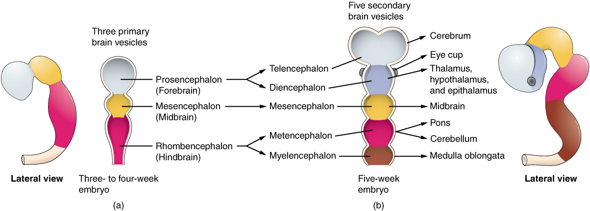
The neural tube develops three major vesicles at a certain stage. I will first provide the developmental names, but I will also include names that are easier to remember. They are the prosencephalon, also known as the forebrain, the mesencephalon, also known as the midbrain, and the rhombencephalon, also known as the hindbrain (19). The prosencephalon has two subdivisions. They are the telencephalon, which later develops into the cerebral hemispheres, and the diencephalon. The mesencephalon, which is the midbrain, has no subdivision. And the rhombencephalon, which is the hindbrain, has like the forebrain two subdivisions. They are the metencephalon, which later develops into the pons and cerebellum, and the myelencephalon, which later becomes the medulla oblongata. The part of the neural tube without the vesicles later becomes the spinal cord.
Side Note
I want to go into more detail about how the same body parts have different names depending whether they are in the CNS or PNS. For example, a collection of cells in the PNS is known as a ganglion, but it is called a nucleus in the central nervous system. However, there are exceptions like the basal ganglia, which is located in the central nervous system. Another example would be tract and nerve. They are basically the same thing, nevertheless, the name of the former is different because it is located in the CNS, while the nerve is located in the PNS. Keep in mind that this happens with other parts.
The Spinal Cord
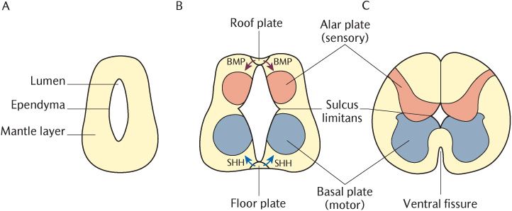
Once the neural tube closes, the spinal cord starts to form. Then, a process called lamination starts to occur. It has this name because it obtains three layers. They are the marginal, the mantle, and the ependymal layer. The first one is filled with white matter, it looks like that because of the collection of axons present there. The second mantle has a darker appearance and it is known as gray matter. The last layer is the ependymal one. This is a layer made up of epithelial cells, which are glial cells that create the epithelial lining of the "brain and the central canal of the spinal cord"(21).
Then, when the neural crest stops developing, we are left with the peripheral nervous system. Like mentioned before this includes the Schwann cells, which are the glial cells that provide the myelin sheath to neurons (remember that the ones in the CNS are called oligodendrocytes). This also includes things outside the spinal cord like the dorsal root ganglion cell, but we will cover more of this later.
In the mantle layer, which can be observed in the picture above, there are two zones where there is a rapid increase of cells. These two zones are called the alar and basal plate (both can also be observed in the picture above). Both plates become the dorsal and ventral horn, respectively. The horns can be seen in the picture down below. The horns are in charge of moving information. The dorsal horn moves sensory information up to the brain, and the ventral horn moves motor info down the spinal cord. The sulcus limitans is a groove that separates the basal and the alar plates.
Resources
1. "Neuroanatomy Text and Atlas" by John H. Martin
2. http://www.ncbi.nlm.nih.gov/books/NBK10869/
3. http://www.dictionary.com/browse/phagocytize
4. http://www.thefreedictionary.com/phago-
5. http://medical-dictionary.thefreedictionary.com/-cyte
6. http://www.dictionary.com/browse/peripheral
7. http://medical-dictionary.thefreedictionary.com/somatic+nervous+system
8. http://www.merriam-webster.com/dictionary/soma
9. http://www.merriam-webster.com/dictionary/autonomic%20nervous%20system
10. http://www.medicinenet.com/script/main/art.asp?articlekey=3286
11. https://www.britannica.com/science/human-nervous-system/The-autonomic-nervous-system#ref942317
12. https://www.sciencedaily.com/terms/parasympathetic_nervous_system.htm
13. http://medical-dictionary.thefreedictionary.com/neural+plate
14. http://www.ncbi.nlm.nih.gov/books/NBK10065/
15. http://www.ncbi.nlm.nih.gov/books/NBK10080/
16. http://www.columbia.edu/cu/psychology/courses/1010/mangels/neuro/navigation/navigation.html
17. http://www.cdc.gov/ncbddd/birthdefects/anencephaly.html
18. http://www.cdc.gov/ncbddd/spinabifida/facts.html
19. http://www.med.umich.edu/lrc/coursepages/m1/embryology/embryo/08nervoussystem.htm
20. http://www.ncbi.nlm.nih.gov/pmc/articles/PMC2799634/
21. https://www.britannica.com/science/ependymal-cell


Comments
Post a Comment