Supporting Cells - Introduction to Psychobiology (Part 3)
Anterograde and Retrograde Axoplasmic Transport
For more information about biopsychology, please check out "Physiology of Behavior" by Neil Carson
Before delving into the topic of supporting cells, I want to cover axoplasmic transport, which was not covered in the second part of this series (http://hbookreviews.blogspot.com/2016/04/introduction-to-psychobiology-part-2.html). It occurs on microtubules, which are a bundle of protein filaments with two main functions (1). One of them is to form the cytoskeleton, which gives each neuron its shape. The other function is to engage in axoplasmic transport. This is the process in which substances are transported along the axon. Kinesin and dynein, both proteins, carry them. If the movement is from the body of the cell towards the terminal buttons, the process is called anterograde and if it is from the terminal buttons to the soma it's called retrograde transport (Antero- means towards and retro- means backwards).
Supporting Cells
Neurons are not the only type of cells in the brain. There are cells, such as glial cells, that support neurons in different ways. For example, the type of cell mentioned above holds neurons together (glia means glue) and provides nutrients (2). There are three main types of glial cells: astrocytes, oligodendrocytes, and microglia (3). The first cell received its name because of its shape (astrocyte means star cell) and it is responsible for delivering nutrients, as well as controlling the development of neurons (4). Another function is to engage in a process known as phagocytosis in which astrocytes engulf and eat cells that have died. The second type of cells, which are oligodendrocytes, produce an insulating sheath called myelin that covers the axons of neurons (5). This facilitates the electricity traveling the axon to engage in exocytosis, but we will cover this process more in detail later on. Finally, it is important to note that the points at which the axon is not insulated are called nodes of Ranvier.

The last type of supporting cell is microglia. They also engage in phagocytosis and act as one of the parts of the immune system in the CNS (6). So far we have only covered the CNS. However, there are supporting cells in the peripheral nervous system (PNS) too such as Schwann cells. They perform the same functions as oligodendrocytes cells (7) from the CNS (remember that the central nervous system is encompassed by the brain and the spinal cord and the PNS is everything outside of it).
The Blood Brain Barrier
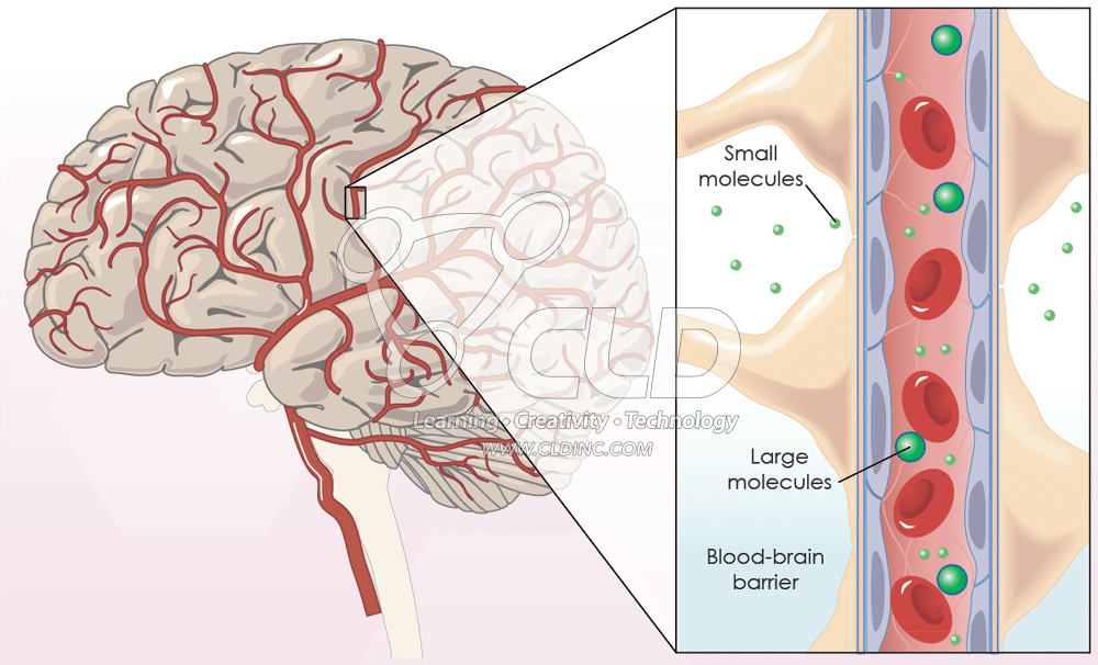 Another important component to mention is the blood brain barrier (BBB). This refers to the barricade that separates the brain and the vascular system (8). Moreover, the BBB is semipermeable, this means that some substances are able to pass through it (2). In this case, the barrier has some gaps that allow substances to come in and out of the vascular system. Another part, besides the small gaps, where substances can travel through the barrier is the areapostrema, which is the section responsible for vomiting. In this area, neurons can detect toxins in the blood and thus induce vomiting (9).
Feel free to leave comments, questions, concerns, or suggestions.
References
1. http://www.ruf.rice.edu/~bioslabs/studies/invertebrates/microtubules.html
2. "Physiology of Behavior" by Neil Carson.
3. http://www.ncbi.nlm.nih.gov/books/NBK10869/
4. http://www.networkglia.eu/en/astrocytes
5. http://www.britannica.com/science/oligodendrocyte
6. http://www.networkglia.eu/en/microglia
7. http://www.britannica.com/science/Schwann-cell


Comments
Post a Comment